Aortic Dissection
Type B Aortic Dissection — Endovascular Treatment
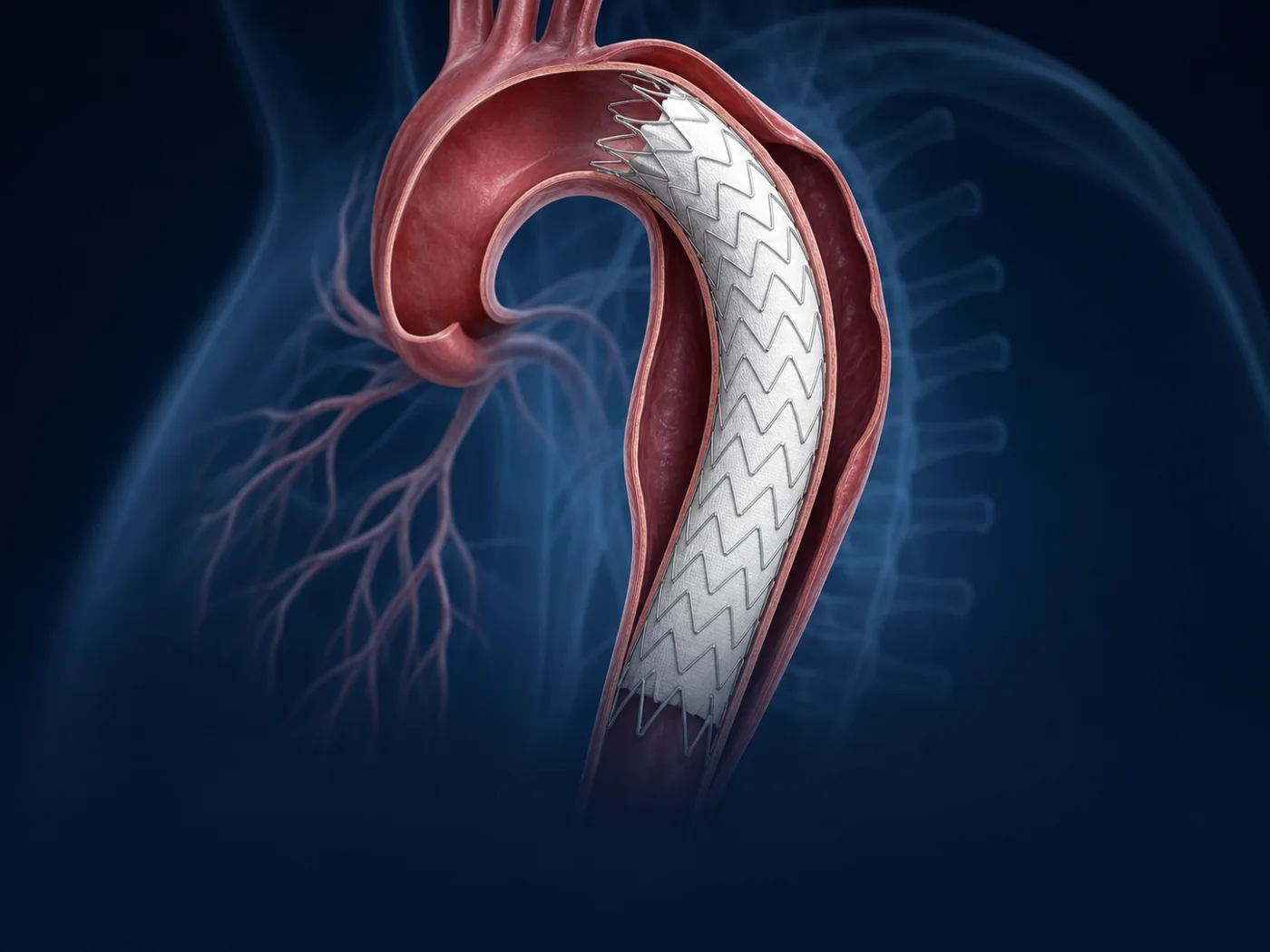
Stent-graft treatment of dissection in the descending aorta.
Overview
What it is
Endovascular treatment of type B dissection uses a stent-graft to cover the entry tear in the descending aorta, redirecting blood flow into the true channel. It is used both in emergencies (complicated dissection) and electively to prevent later aneurysm growth and complications.
When it’s recommended
- Complicated type B dissection (malperfusion, rupture, pain)
- Selected uncomplicated dissections at risk of later growth
- Anatomy suitable for stent-graft coverage
How it’s performed
Every plan is individualized and reviewed by our multidisciplinary aortic team.
Urgent or planned CT angiography
A stent-graft covers the primary entry tear
Flow is redirected to the true lumen, branches preserved
Close imaging follow-up tracks aortic remodeling
Recovery
Recovery & follow-up
Recovery depends on whether treatment is urgent or elective; all patients enter a structured surveillance imaging program.
Understanding aortic dissection
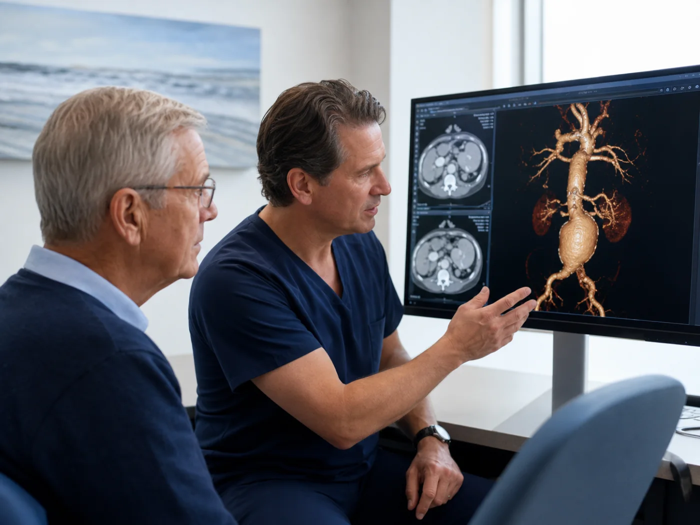
An aortic dissection begins when the inner lining of the aorta tears, letting blood enter the wall itself and split it into two channels — the original (true) channel and a new (false) channel. This can reduce blood flow to vital organs, weaken the aorta, and, if untreated, lead to rupture.
Types and timing
Physicians describe a dissection by where it starts and how long ago it happened:
- Type A involves the ascending aorta near the heart and is usually a surgical emergency.
- Type B begins beyond the arch branches — often managed with medication when uncomplicated, or with endovascular repair when complicated by rupture or reduced blood flow (malperfusion).
- Timing ranges from acute (within two weeks) to subacute and chronic (beyond 90 days), which shapes how the aorta is treated.
Aortic dissection affects roughly 10–15 people per 100,000 each year and is 2–5 times more common in men. About a third are Type B; most are uncomplicated at first, but lifelong surveillance is essential.
Who is at risk
- Long-standing high blood pressure
- Smoking and stimulant use
- Genetic and connective-tissue conditions such as Marfan, Loeys-Dietz, and vascular Ehlers-Danlos syndromes
- A bicuspid aortic valve or a family history of aortic disease
Minimally invasive treatment & evidence
For complicated Type B dissection, national guidelines (ACC/AHA, 2022) recommend minimally invasive endovascular repair over open surgery when the anatomy is suitable. In selected patients with chronic dissection, the true channel can become too narrow to safely place a stent-graft. Dr. Oderich helped develop transcatheter electrosurgical septotomy (TES) — a catheter-based technique that uses a fine electrified wire to divide the membrane between the two channels, reopening space for a durable minimally invasive repair.
13 → 28 mm
Average true-channel width, before → after TES
Kanamori et al., J Vasc Surg 2024
~3{a67e48988fb40e36bdb74a4903c6bdee865f13ce06fb3094ced242c5bab80628}
30-day mortality
121-patient series · Figueroa et al., J Vasc Surg 2025
93{a67e48988fb40e36bdb74a4903c6bdee865f13ce06fb3094ced242c5bab80628}
Freedom from seal-zone reintervention
Figueroa et al., J Vasc Surg 2025
Figures reflect published multicenter series and registries — not a promise of individual results, which vary with each patient’s anatomy and overall health.
Why the Baylor Medicine Center for Aortic Surgery
Complex aortic care is safest in experienced hands. Dr. Oderich and the Center bring more than 7,000 open and endovascular aortic repairs of experience, advanced imaging, and a multidisciplinary team to every case.